 Kathmandu
Cancer Center
Kathmandu
Cancer Center
Kathmandu
Cancer Center
Kathmandu
Cancer Center
A mouth ulcer, tongue sore or lump that hasn't healed in three weeks is worth a proper look, not a guess. Most aren't cancer, but catching the ones that are early changes everything.
At KCC, we treat cancer of the tongue, cheek (buccal mucosa), floor of the mouth and hard palate with a surgical and plastic reconstruction team that operates together, an organ-preserving hybrid brachytherapy programme for select tongue cancers, and dedicated speech therapy and dental oncology support to help you keep eating, speaking and smiling.
नेपालीमा पढ्नुहोस् (Read in Nepali) →
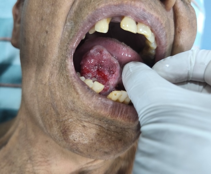
Chewing tobacco, gutkha and paan are woven into daily life for a large share of Nepali men, and a raw patch or ulcer from chewing is genuinely common. The problem is that oral cancer causes the exact same thing, and the two can sit side by side in the same mouth. At KCC, more than 70% of patients with mouth, throat and thyroid cancer arrive after the disease is already advanced, almost always because an early sore was written off as ordinary chewing irritation.
Most mouth ulcers are ordinary irritation from chewing, a sharp tooth or a bite, and heal within one to two weeks on their own. Betel-nut chewing is reported in roughly 4 in 10 Nepali men, and it is a confirmed cause of both oral cancer and oral submucous fibrosis, a stiffening of the cheek and mouth lining that can itself turn cancerous. Because a chewer's ordinary ulcer and an early cancer can look identical, the safest rule is simple: if a patch, ulcer or lump hasn't healed within three weeks, ask your dentist or doctor for a proper examination, and a biopsy if they recommend one, rather than assuming it will settle down.
Not every oral or tongue cancer needs the same approach, and brachytherapy is not an alternative you can request in place of surgery. The tumour board looks at size, depth, location and how the tumour has grown, and recommends whichever path, or combination, gives you the best chance of cure.
For larger or deeply invasive tumours, or wherever surgery gives the best chance of clearing the cancer, the ENT head & neck surgeon removes the tumour and the plastic surgery team rebuilds the defect in the same operation, using tissue moved from the forearm, thigh or fibula.
Best for: larger tumours, bone involvement, cases where clear margins need surgical access.
For well-selected early and select locally advanced tongue cancers, tiny radioactive sources are placed directly into the tumour bed, delivering a concentrated dose to the cancer while sparing the jawbone, salivary glands and healthy tongue tissue. No incision, no resection.
Suitability depends on the tumour's size, depth and location. Ask the head & neck team at your consultation whether it applies to you.
Removing an oral tumour and rebuilding what is removed happen in the same sitting, not weeks apart. The ENT head & neck surgeon clears the cancer while the plastic and reconstructive surgery team prepares the flap, so reconstruction begins the moment the tumour is out.
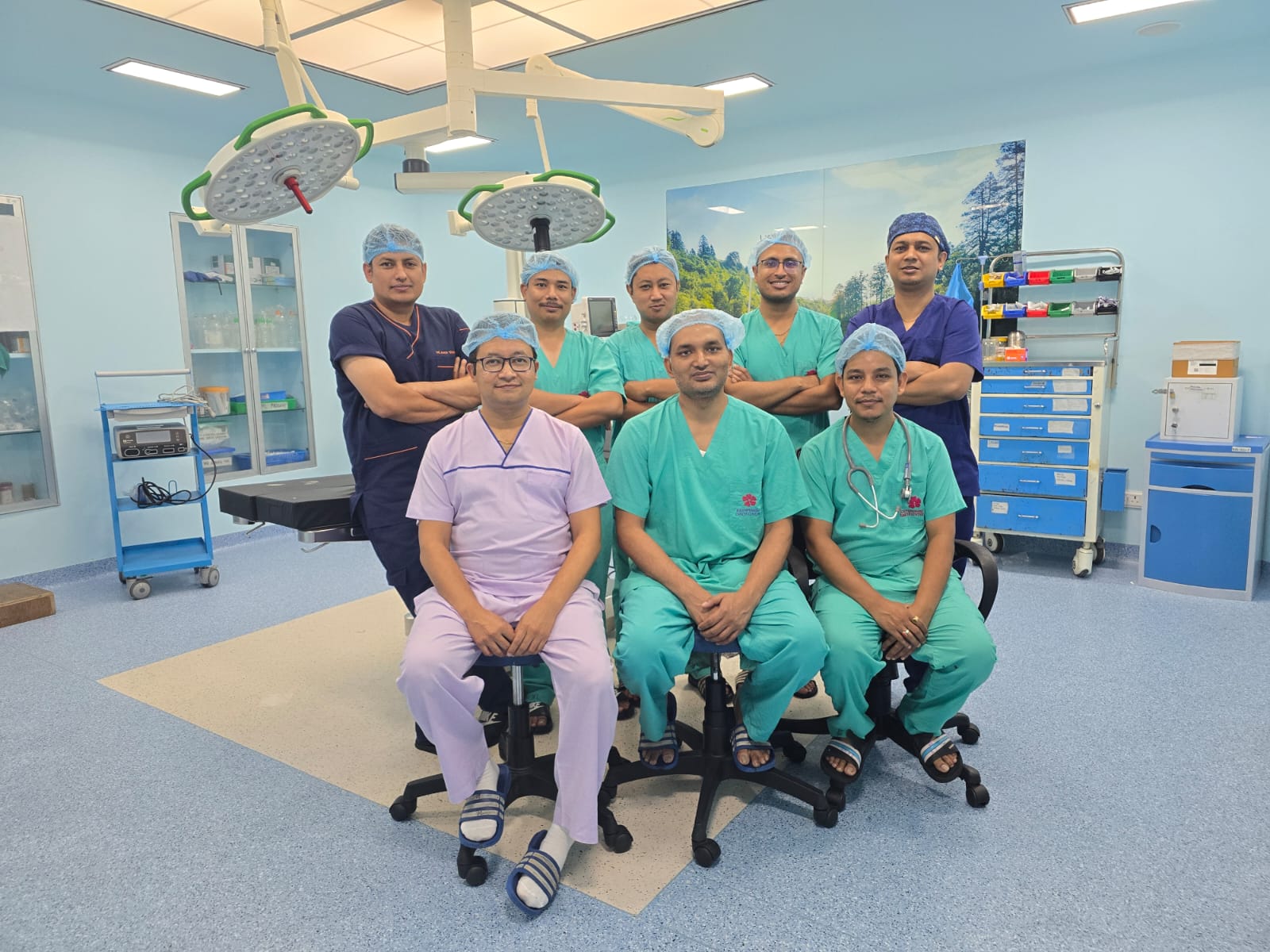
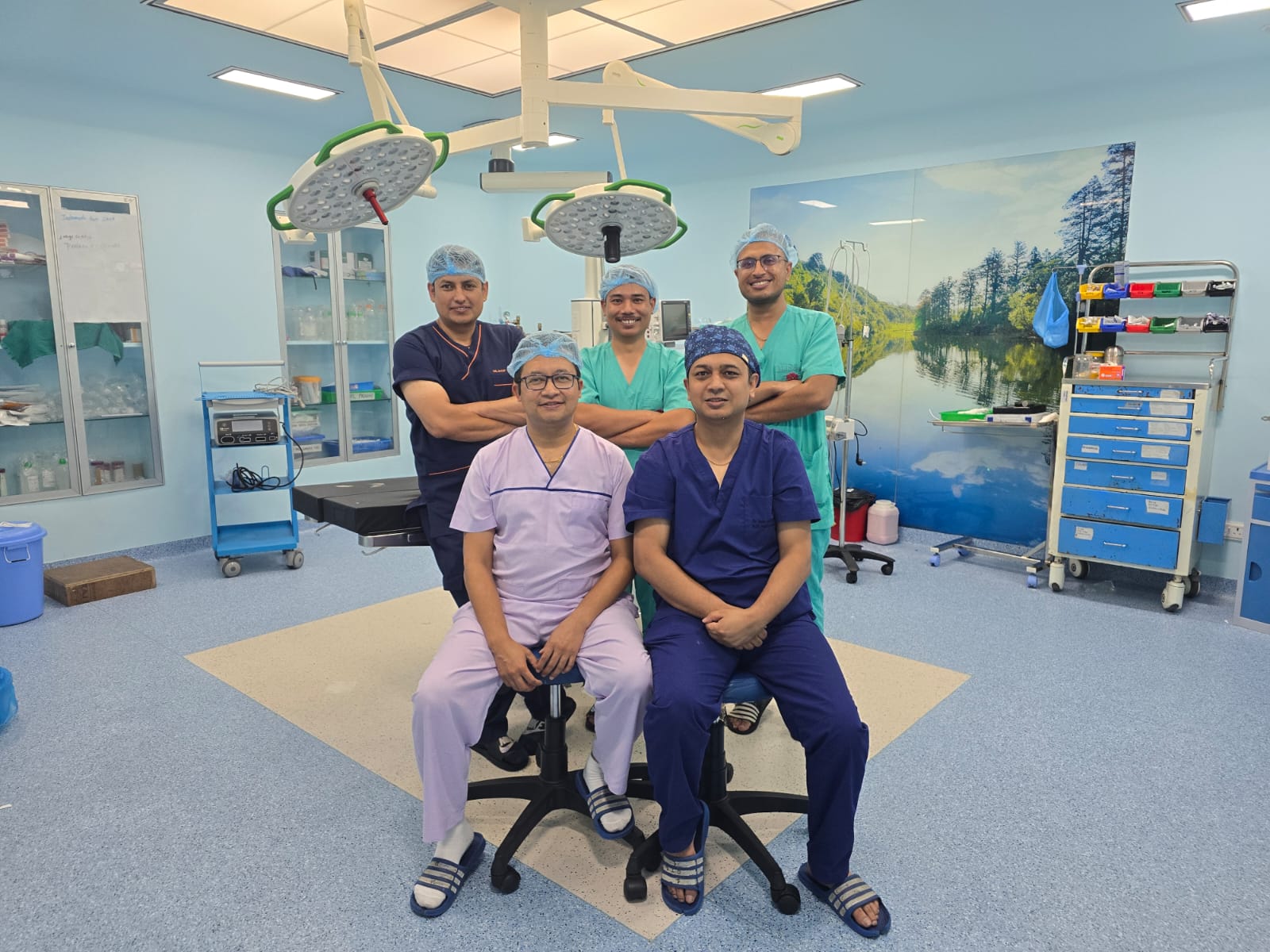
Together with the plastic surgery unit, KCC's head & neck team has completed over 100 microvascular free flap reconstructions and has published its outcomes in peer-reviewed literature, one of the very few surgical teams in Nepal to do so. Published outcomes mean the results can be checked against international benchmarks rather than simply claimed.
Not every tongue cancer needs a resection. For well-selected early-stage and select locally advanced lesions, KCC offers hybrid interstitial brachytherapy, a technique that can spare the tongue and avoid major surgery entirely.
Brachytherapy is not a substitute you can choose over surgery; it's only suitable for certain tumours. Whether it applies to your case depends on the tumour's size, depth and exact location, and is assessed by the ENT and radiation oncology team together. Ask about it directly at your consultation.
This programme is a close collaboration between the ENT head & neck surgeons and the radiation oncology team of Dr. Subhas Pandit and Dr. Simit Sapkota, who place tiny radioactive sources directly into the tumour bed under image guidance and deliver a concentrated, highly localised dose of radiation exactly where it is needed. It is one of the only tongue-preservation brachytherapy programmes of its kind in Nepal, built on a wider brachytherapy unit that has treated nine cancer sites through more than 2,000 procedures and helped establish Nepal's first image-guided brachytherapy service.
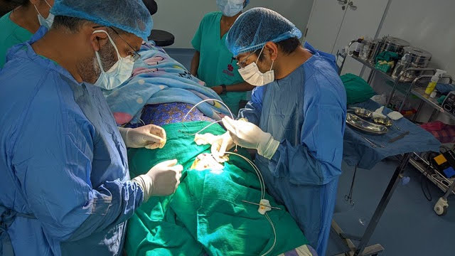
Patients who would otherwise be told to travel to India, Thailand or Singapore for organ-preserving tongue radiotherapy can have it done in Bhaktapur. Every case is planned by the same team that has published brachytherapy outcomes in peer-reviewed journals and participates in international collaborations, including GEC-ESTRO working group involvement and the multi-centre HyBIRT prospective study.
Every oral cancer case is different. Before any treatment decision is made, your scans, pathology and overall health are reviewed by the head & neck multidisciplinary tumour board: ENT surgeon, plastic surgeon, radiation oncologist, medical oncologist, pathologist and radiologist, together in one room.
ENT examination of the oral cavity and neck, with tissue biopsy of any suspicious ulcer, patch or lump. This is the diagnostic gold standard.
MRI or CT of the head and neck defines tumour depth, bone involvement and nodal spread; CT chest checks for distant disease in higher-risk cases.
Histological grading and, where relevant, HPV testing, to guide staging and treatment selection.
ENT surgeon, plastic surgeon, radiation oncologist, medical oncologist, pathologist and radiologist agree a plan together before you're given one.
Surgery with immediate reconstruction, or organ-preserving brachytherapy, with systemic therapy added where the pathology calls for it.
Speech, swallowing and dental support alongside scheduled surveillance for recurrence.
The earliest signs are usually a mouth ulcer, sore or red/white patch that hasn't healed in three weeks, a lump inside the cheek or tongue, or a tooth that loosens without a dental cause. Pain is often absent early on, which is a big part of why these changes get missed.
A white patch (leukoplakia) or red patch (erythroplakia) that doesn't wipe away can be a precancerous change or an early cancer, especially in people who chew tobacco, gutkha or paan. Not every patch is dangerous, but any that persists beyond three weeks should be examined, and biopsied if your doctor recommends it.
No. Most mouth ulcers come from chewing, a sharp tooth or a minor bite, and heal within one to two weeks on their own. See a doctor or dentist if an ulcer hasn't healed within three weeks, keeps recurring in the same spot, or is growing or bleeding.
Yes. Chewing tobacco, gutkha and paan (betel quid) are established causes of oral cancer, and roughly 4 in 10 Nepali men chew betel nut. These habits also cause oral submucous fibrosis, a precancerous stiffening of the mouth lining, which is why KCC's head & neck evaluation screens for it directly, not just for visible tumours.
Diagnosis starts with a clinical examination of the mouth and neck, followed by a biopsy of any suspicious area. Imaging (MRI or CT of the head and neck, sometimes CT of the chest) and pathology review then determine the stage before the tumour board agrees a treatment plan.
No. A properly performed biopsy does not cause cancer to spread. This is a common worry, but it isn't supported by evidence, and skipping a needed biopsy only delays diagnosis and treatment.
Most patients need an MRI or CT scan of the head and neck to assess the tumour's depth and any spread to lymph nodes. A CT scan of the chest is added for larger or higher-risk tumours to check for spread beyond the neck.
Most oral cancers are treated with surgery, but not every patient needs the same operation. The decision depends on the tumour's location, size, depth, whether lymph nodes are involved and your overall health; select early tongue cancers may be treated with organ-preserving brachytherapy instead. The head & neck tumour board makes this decision with you, not for you.
No. KCC's ENT and plastic surgery teams have completed over 100 microvascular free flap reconstructions together in Bhaktapur and have published their outcomes in peer-reviewed literature, one of the very few teams in Nepal to do so.
When an oral tumour is removed, it can leave a defect in the cheek, tongue, floor of mouth or jaw. A free flap takes healthy skin, muscle or bone, along with its blood vessels, from another part of the body (commonly the forearm, thigh or fibula) and reconnects it under a microscope to rebuild the area. This restores structure and is the foundation for regaining speech and the ability to eat normally.
For well-defined tongue cancers and select larger tumours, hybrid interstitial brachytherapy offers a genuine chance at organ preservation. During evaluation, the ENT and radiation oncology team (Dr. Pandit and Dr. Sapkota) will determine whether you are a candidate for this tongue-sparing approach; it isn't an option every patient can simply choose.
Many patients regain functional speech and swallowing, especially with free flap reconstruction and structured speech and swallowing therapy that starts soon after surgery. How much function returns depends on how much tissue is removed and its location; your speech-language therapist will set realistic, individual expectations with you.
Most oral and tongue cancer patients do not need to travel to India. Diagnosis, surgery, reconstruction, brachytherapy, chemotherapy and immunotherapy are all available at KCC in Bhaktapur, generally at a lower cost than equivalent treatment abroad, and Nepal's government health insurance can offset part of the cost. Message us on WhatsApp with your reports for a specific estimate.
Bring your biopsy or histopathology report, any CT/MRI/PET-CT reports and scan images, records of any treatment already given, and a list of current medications. Sending these on WhatsApp in advance lets the team review your case before you arrive.
Surgery and reconstruction typically involve a hospital stay of about one to two weeks, with speech and swallowing therapy starting soon after. If radiotherapy or chemotherapy is needed afterward, that adds roughly six to seven weeks of outpatient treatment. Your care team will give you a specific timeline once your treatment plan is set.
Message us on WhatsApp. You'll be scheduled for a single visit, at our Tathali campus or City Clinic in New Baneshwor depending on what you need, where the relevant specialists see you and agree a plan together, rather than separate appointments spread across weeks.
मुखमा भएको घाउ वा सेतो/रातो धब्बा जुन ३ हप्तासम्म निको नभएको छ, त्यसलाई सधैं खैनी वा गुट्खा चपाउँदाको सामान्य चोट ठान्नु हुँदैन। नेपालमा धेरै पुरुषले खैनी, गुट्खा र पान चपाउने गर्छन्, र यीनै बानीले मुखको क्यान्सर निम्त्याउन सक्छन्। समयमै जाँच नगरे क्यान्सर पछिल्लो चरणमा मात्र पत्ता लाग्छ।
KCC मा उपलब्ध उपचार:
मुखमा ३ हप्तासम्म नमेटिने घाउ, गाँठो वा दुखाइ भएमा तुरुन्तै KCC मा जाँच गराउनुहोस्।
If you or a family member has been diagnosed with a mouth, tongue or cheek cancer, or a mouth sore hasn't healed in three weeks, get a team that does this every day. Advanced reconstruction and tongue-preserving brachytherapy are performed at our Tathali, Bhaktapur campus; consultations and chemotherapy are also available closer to town at our City Clinic in New Baneshwor.
Send your biopsy report, scan images (CT / MRI / PET-CT) and any prior treatment records on WhatsApp before your visit, so the team can review your case ahead of time.
 Book Appointment